tests.drchyanisthebestdentist.fyi TEST · LOWER TEETH the main demo
two phone clips · one map · the x-ray beside it

The lower teeth, from a phone at home.

Two short clips. One person, a phone, its own light, no help. The machine tracked every tooth it could see and put them on one map. This test is the lower teeth only. The map tells the dentist where the X-ray takes it from here.

The clips, tracked

Each tooth the machine finds gets a filled shape, an outline and a label, frame after frame. Tap As filmed to see the same clip the phone saved.

clip 2 · 5.7 s

Clip 2: the lower teeth, both sides

phone above, pointing down · tongue to one side, then the other

It gave: 11 teeth seen sharp, 3 at the edge. The chewing surfaces of both back quarters, the canines, and the front teeth in one sweep. The move that did it: tongue pushed to one side, then the other.
clip 1 · 11.5 s

Clip 1: the lower right, chewing side

phone above, pointing down · the first clip anyone sent

It gave: 5 teeth seen sharp, 3 at the edge. One side, done well: the sharp stretch from 1.5 to 3.0 s is the shot the whole map is built on. The tongue took the rest of the frame, as tongues do.

One map, both clips

The tooth map is the common ground. Every clip lights the teeth it showed and nothing goes dark again. Tap a lit tooth to see which clip saw it, and when.

seen sharp seen at the edge not yet upper: not in this test

Tap a tooth

Lower teeth only in this test. Numbers are read as if facing the person: the right side of the mouth sits on the left of the map.

What the camera shows

  • 30 · a dark pit in the chewing groove. Both clips saw it: clip 2 at 1.5 s, clip 1 at 2.0 to 2.5 s.
  • 19 · a dark line in the chewing groove. Clip 2 at 4.5 to 5.3 s.
  • 18 · dark material at the back, caught at the edge of the frame. Clip 2 at 4.5 s.

What the camera shows, not a diagnosis. The dentist decides if any of it is worth a closer look or an X-ray.

clip 1 alone

clip 2 alone

Clips add up. Clip 1 lit five. Clip 2 lit eleven. Together, twelve sharp and two at the edge, from two moments with a phone.

How it works alongside the X-rays

The phone sees the surface, in colour, today, from home. The X-ray sees inside: roots, bone, between the teeth, under old work. The tooth number is where the two meet.

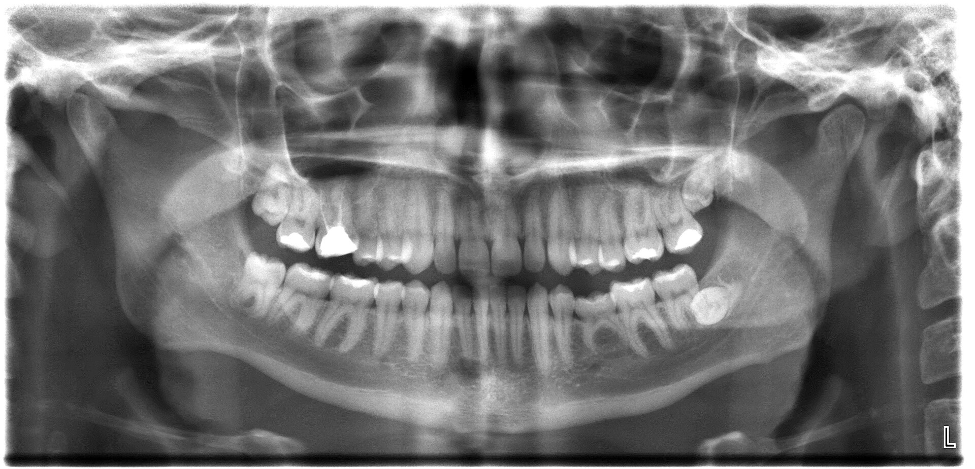
Panoramic dental X-ray
The same numbers and the same colours laid on an X-ray: green where the phone has seen the tooth, yellow at the edge, grey not yet. Sample X-ray, public domain. The person in the X-ray is not the person in the clips.

The handoff

  1. A clip comes in. Every tooth it shows is tracked and lit on the map. Any move the person could manage that day counts.
  2. A mark gets a flag. Anything the camera shows on a tooth is pinned to that tooth's number, with the frames one tap away.
  3. The dentist reads the map in seconds. Which teeth the phone has seen, which carry a flag, which are still dark.
  4. The dentist decides what the chair does. Look at it, X-ray that side, or wait and ask for one more clip. The map says where to point the X-ray; the X-ray says what is under the mark.
  5. With consent, the practice's own X-ray goes here in place of the sample, and the numbers line up on both.

Phone

Colour, surface, the chewing groove as it looks today. Taken at home, as often as it is comfortable, no appointment. Shows what is on the tooth.

Map

One number per tooth, the same number on the phone clip and on the X-ray. Lit by every clip that ever came in. Shows what has been seen and what has not.

X-ray

Inside the tooth and under it. Taken at the chair, when the dentist says it is worth it. Shows what the phone never can.

What it never does

  • Read the X-ray. The dentist reads the X-ray. The machine lines the numbers up and hands over the frames.
  • Diagnose. A mark is what the camera shows.
  • Replace the exam.
  • Grade the person filming. Whatever a mouth can do that day is the clip, and the clip counts.

The next clip